Bones In Your Leg Diagram - Leg pain can also be caused by blood clots, varicose veins or poor circulation.. This keeps the bones together, giving a high ankle sprain time to heal. Pain in the large bones and muscles of the upper leg can be caused by a wide range of ailments. The bones of your leg and foot helped give you the ability to score that field goal. This allows weight to be distributed either anteriorly or posteriorly throughout the foot. The second metatarsal bone is the longest.
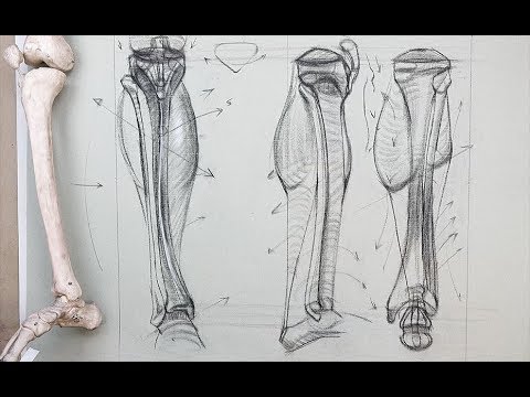
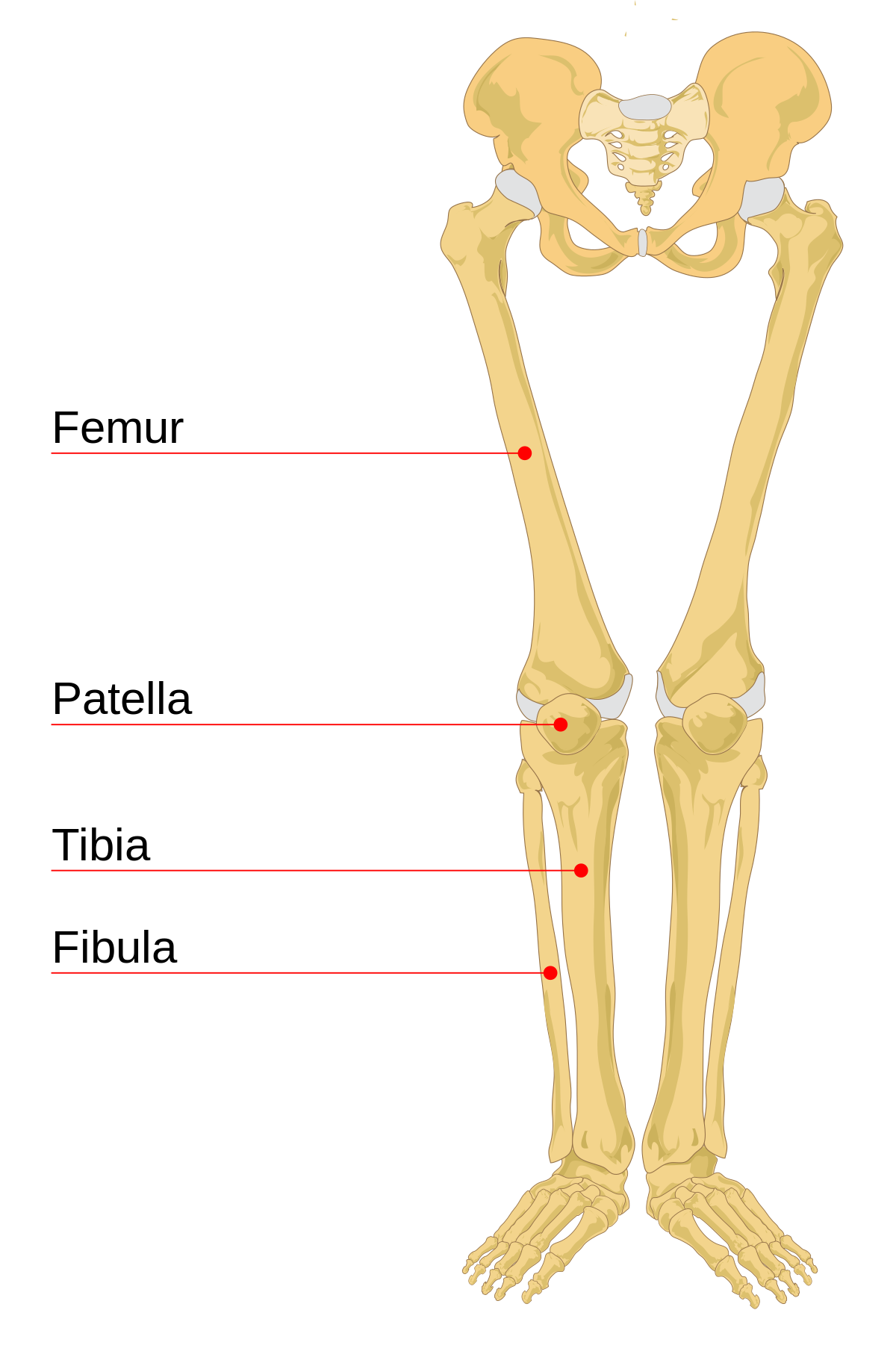
In humans the neck of the femur connects the shaft and head at a 125 degree angle, which is efficient for walking. The smaller bone that runs alongside the tibia (fibula) and the kneecap (patella) are the other bones that make the knee joint. The bones of the leg are the femur, tibia, fibula and patella.the foot bones shown in this diagram are the talus, navicular, cuneiform, cuboid, metatarsals and calcaneus. The major bones of the leg are the femur (thigh bone), tibia (shin bone), and adjacent fibula, and these are all long bones.the patella (kneecap) is the sesamoid bone in front of the knee.most of the leg skeleton has bony prominences and margins that can be palpated and some serve as anatomical landmarks that define the extent of the leg. The human leg consists of 8 bones, 4 per leg.
Lower Leg Bone Anatomy Anatomy Drawing Diagram from i.ytimg.com Some common causes of leg pain include: The bones of your leg and foot helped give you the ability to score that field goal. Ankle & lower leg anatomy. Your hamstring is directly connected to your ischium bones, and any tear or damage to your hamstring can result in sit bone pain. Distal to the ankle is the foot. The bones of the hip include the femur, the ilium, the ischium, and the pubis. In humans the neck of the femur connects the shaft and head at a 125 degree angle, which is efficient for walking. The knee joint is the largest joint in the body and is primarily a hinge joint, although some sliding and rotation occur.
Muscle anatomy gluteus 12 photos of the muscle anatomy gluteus gluteus muscle anatomy ct, gluteus muscle anatomy mri, human muscle anatomy gluteus maximus, muscle anatomy gluteus, muscle anatomy of gluteal, human muscles, gluteus muscle anatomy ct, gluteus muscle anatomy mri, human muscle anatomy gluteus maximus.
Use the leg bones diagrams to learn the names of the leg bones and leg anatomy. Lower limb, 3d scan, angiography scanner 3d of the right calf, visualization of the skeleton system, tibia and fibula, and vascularization of the. The journal of manual and manipulative therapy reports that hamstring injuries can cause buttock pain on the side where the injury occurred. Also called the shin bone, the tibia is the longer of the two bones in the. The pubis, ischium, and ilium together constitute the pelvis while the thigh bone is the femur. The smaller lateral bone of the lower leg. Its lower end helps create the knee joint. This keeps the bones together, giving a high ankle sprain time to heal. The knee joint is the largest joint in the body and is primarily a hinge joint, although some sliding and rotation occur. In popular usage, the leg extends from the top of the thigh down to the foot. The smaller bone that runs alongside the tibia (fibula) and the kneecap (patella) are the other bones that make the knee joint. The second metatarsal bone is the longest. The sacrum and the coccyx attach to the two hip bones to form the pelvis, but are more important to the spinal column, where they are counted.
This area is commonly referred to as the calf. The proximal portion of the tibia is tibial plateau which acts as a cusp for the knee, the distal portion tapers into the medial malleoli and the concave surface which articulates with the talus at the ankle joint. Many muscles that move the trunk and legs, such as our abdominal muscles, attach to the hip bones. The leg is specifically the region between the knee joint and the ankle joint. This keeps the bones together, giving a high ankle sprain time to heal.
Pin On Anatomy from i.pinimg.com The smaller bone that runs alongside the tibia (fibula) and the kneecap (patella) are the other bones that make the knee joint. #diagram and names of leg bones #diagram of foot and leg bones #diagram of leg bones #diagram of lower leg bones #diagram of the bones in your leg related posts of diagram of leg bones long bone femur label The lower leg is comprised of two bones, the tibia and the smaller fibula. The major bones of the leg are the femur (thigh bone), tibia (shin bone), and adjacent fibula, and these are all long bones.the patella (kneecap) is the sesamoid bone in front of the knee.most of the leg skeleton has bony prominences and margins that can be palpated and some serve as anatomical landmarks that define the extent of the leg. Some types of leg pain can be traced to problems in your lower spine. There are a total of 60 bones in the legs. Treatment of a broken leg depends on the location and severity of the injury. However, in medical terminology, the leg refers to the portion of the lower extremity from the knee to the ankle.
At the same time, the bones and joints of the leg and foot must be strong enough to support the body's weight while remaining.
The knee joint is the largest joint in the body and is primarily a hinge joint, although some sliding and rotation occur. The bones together make up the hip. They are numbered from one to five, starting from the medial (inner) side of the foot. These landmarks are the anterior superior iliac spine. He leg's main function in the human is for locomotion and support of the rest of the body. Some types of leg pain can be traced to problems in your lower spine. Muscle anatomy gluteus 12 photos of the muscle anatomy gluteus gluteus muscle anatomy ct, gluteus muscle anatomy mri, human muscle anatomy gluteus maximus, muscle anatomy gluteus, muscle anatomy of gluteal, human muscles, gluteus muscle anatomy ct, gluteus muscle anatomy mri, human muscle anatomy gluteus maximus. The tibia and the fibula. The leg is specifically the region between the knee joint and the ankle joint. Related posts of muscles and tendons of the leg muscle anatomy gluteus. Take a look at the diagram of foot bones below. The tibia and fibula are two long bones that run parallel to each other, forming the scaffold of the leg and providing attachment points for many muscles. A severely broken leg may require surgery to implant devices into the broken bone to maintain proper alignment.
Whether treatment is a home remedy or provided by a medical professional, identifying the upper leg pain causes can be useful in determining how to move forward. The leg has two bones: In addition, the broad hip bones provide protection to the delicate internal organs of the pelvis, such as the intestines, urinary bladder, and uterus. The second metatarsal bone is the longest. Some types of leg pain can be traced to problems in your lower spine.
Leg Bone Wikipedia from upload.wikimedia.org The smaller bone that runs alongside the tibia (fibula) and the kneecap (patella) are the other bones that make the knee joint. However, in medical terminology, the leg refers to the portion of the lower extremity from the knee to the ankle. Common causes include falls, motor vehicle accidents and sports injuries. The proximal portion of the tibia is tibial plateau which acts as a cusp for the knee, the distal portion tapers into the medial malleoli and the concave surface which articulates with the talus at the ankle joint. Pain in the large bones and muscles of the upper leg can be caused by a wide range of ailments. This area is commonly referred to as the calf. Let's review all of these bones one last time. The lower leg extends from the knee to the ankle.
Distal to the ankle is the foot.
The hip itself is a ball and socket joint, much like the shoulder.the structures necessary to create this joint are the socket, the joint capsule, muscle, ligaments, and the neck. The leg has two bones: Whether treatment is a home remedy or provided by a medical professional, identifying the upper leg pain causes can be useful in determining how to move forward. The second metatarsal bone is the longest. The five metatarsals are the long bones that link the tarsal bones to the toes, seen in yellow in the diagram below. The human leg consists of 8 bones, 4 per leg. Most leg pain results from wear and tear, overuse, or injuries in joints or bones or in muscles, ligaments, tendons or other soft tissues. The talocrual joint is made up of three main bones. The smaller lateral bone of the lower leg. Browse 7,069 leg bone stock photos and images available, or search for human leg bone or leg bone xray to find more great stock photos and pictures. The smaller bone that runs alongside the tibia (fibula) and the kneecap (patella) are the other bones that make the knee joint. This allows weight to be distributed either anteriorly or posteriorly throughout the foot. Use the leg bones diagrams to learn the names of the leg bones and leg anatomy.
The femur, or thighbone, is the longest and largest bone in the human body leg bones diagram. Your hamstring is directly connected to your ischium bones, and any tear or damage to your hamstring can result in sit bone pain.


0 Komentar